The battle against colorectal cancer (CRC) is entering a new, digital phase. As the global population ages and cancer incidence rises, the pressure on pathology laboratories has reached a breaking point. A recent scientific breakthrough, highlighted by Medical Xpress, introduces a new Artificial Intelligence (AI) model that promises to radically transform how tissue samples are analyzed, offering speed and precision previously thought unattainable.
Colorectal cancer is one of the leading causes of cancer-related deaths worldwide. Early and accurate diagnosis is the most critical factor for patient survival. Traditionally, pathologists examine hundreds of tissue slides under a microscope, searching for subtle morphological changes that indicate the presence of cancer cells, their grade of aggressiveness, and potential response to specific treatments. This process, while the "gold standard," is extremely time-consuming and subject to inter-observer variability.
The Technological Edge of Foundation Models
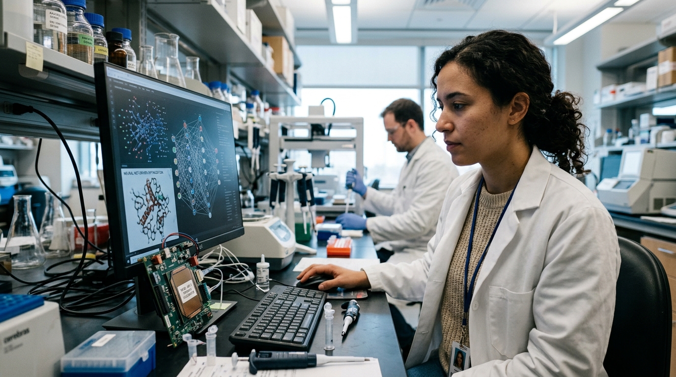
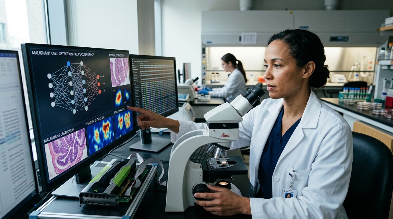
The new AI model is not merely a simple image recognition algorithm. It is built on the architecture of "foundation models," similar to the technology powering Large Language Models (LLMs). Trained on vast databases of digitized histological images, the system has learned to recognize complex patterns that often escape the human eye. The model's ability to process enormous volumes of data in seconds allows for the rapid triage of samples, prioritizing the most critical cases for immediate review.
One of the most impressive features of this technology is its ability to predict the genetic instability of a tumor, such as Microsatellite Instability (MSI). MSI is a crucial biomarker that determines whether a patient will respond to immunotherapy. Until now, determining MSI status required expensive and time-consuming molecular testing. The new AI model can "read" this information directly from a standard biopsy image, significantly reducing costs and waiting times for patients.
From Bench to Bedside: Challenges and Prospects
Despite the impressive performance, integrating AI into daily clinical practice is not without its challenges. The "black box" issue remains central: how can we trust a diagnosis when we do not fully understand the algorithm's decision-making process? Researchers are working feverishly on "Explainable AI" (XAI), ensuring the system can point out the specific tissue areas to the physician that led to its conclusion.
Furthermore, there is the issue of standardization. Tissue samples can vary depending on the staining method or the digitization equipment used by different hospitals. The new model has been designed to be "robust" against these variations, ensuring that its accuracy remains high regardless of the sample's origin. This flexibility is essential for the global adoption of the technology, especially in resource-limited settings.
The Future of Personalized Medicine
The application of AI in colorectal cancer diagnosis is just the beginning. The model's ability to combine histological data with clinical information paves the way for truly personalized medicine. In the future, AI will be able to predict not only the diagnosis but also the likelihood of recurrence or the occurrence of side effects from chemotherapy.
In conclusion, the research presented highlights Artificial Intelligence not as a replacement for the pathologist, but as a powerful co-pilot. The synergy of human expertise and computational power promises to make cancer a more manageable disease, offering hope to millions of patients worldwide. The challenge now shifts from the technical level to the regulatory and ethical spheres, as society is called upon to define the framework within which technology will serve life.