Osteoarthritis (OA) is a primary driver of global disability, affecting millions and costing healthcare systems billions annually. Despite its prevalence, the disease remains a clinical enigma: why do some patients with severe structural damage on MRI feel no pain, while others with seemingly healthy joints experience debilitating agony? New research published on ArXiv (2606.05357) aims to solve this paradox through an innovative AI framework that prioritizes interpretability and trustworthiness.
The Challenge of Longitudinal Research
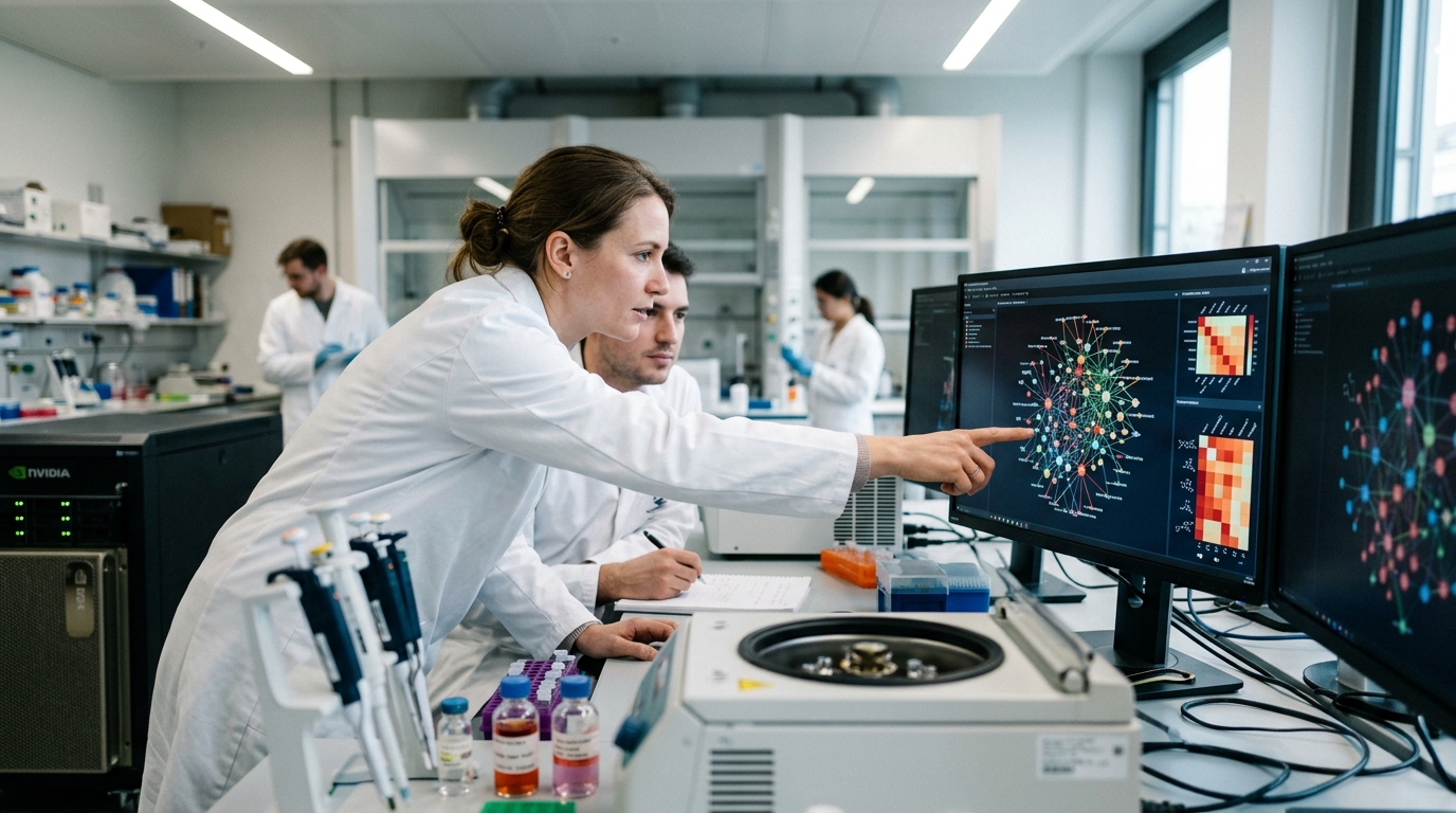
Studying OA requires patience. Changes in cartilage, bone, and meniscus tissue occur slowly over years, not weeks. The Osteoarthritis Initiative (OAI) has amassed a monumental dataset, including thousands of MRIs and clinical pain reports. However, analyzing this data via human radiologists is a Herculean and prohibitively expensive task. This is where Artificial Intelligence steps in, capable of processing vast image libraries in a fraction of the time.
The issue with traditional deep learning models is their "black box" nature. They provide predictions without explanation. In medicine, where decisions directly impact human lives, blind reliance on an algorithm is a liability. The new framework proposed by the researchers addresses this by merging the raw power of deep learning with the rigor of interpretable statistical modeling.
Automating the MOAKS System
At the heart of this system is the automated prediction of the MRI Osteoarthritis Knee Score (MOAKS). MOAKS is a standardized scoring system that evaluates various structural features of the knee, such as bone marrow lesions (BMLs), cartilage loss, and meniscus tears. Manually scoring a single MRI can take an expert radiologist up to an hour.
The proposed AI model was trained to identify these features with accuracy nearing that of a specialist. Utilizing Convolutional Neural Networks (CNNs), the system maps the knee joint and pinpoints pathological areas. This enables large-scale studies involving thousands of subjects—a feat previously impossible. The innovation, however, lies not just in the speed of analysis, but in how this structural data is linked to patient-reported outcomes.
Statistical Interpretability and Trust
Instead of attempting to predict pain directly from raw pixels—an approach often fraught with error due to the subjective nature of pain—the framework uses predicted MOAKS scores as intermediate variables in a statistical model. This model analyzes how structural changes (e.g., an increase in bone marrow lesions) correlate with pain progression over time.
This approach offers two distinct advantages. First, it is interpretable: clinicians can see exactly which structural changes are driving the pain prediction. Second, it is trustworthy, as it relies on clinically validated biomarkers. The study demonstrates that certain lesions are more closely associated with pain than others, confirming clinical suspicions with the statistical weight of big data.
The Future of Personalized Care
The implications of this framework extend far beyond academic research. In the future, such a system could be integrated into clinical practice, helping orthopedists identify patients at the highest risk of rapid progression. Furthermore, it could revolutionize clinical trials for new OA drugs by providing an objective, rapid measurement of treatment efficacy.
In conclusion, this work represents a significant leap toward "intelligent" radiology. It proves that AI does not have to be an opaque puzzle; it can be a transparent and powerful ally in understanding the most complex ailments of the human body. The challenge now shifts to validating these models across diverse populations and integrating them into the daily workflows of modern hospitals.