In the high-stakes world of modern anesthesiology, the "difficult airway" remains a primary concern for clinicians. It is a scenario where a skilled anesthesiologist encounters significant challenges in mask ventilation, tracheal intubation, or both. Despite decades of clinical evolution, traditional predictive methods—such as the Mallampati score, thyromental distance, and neck mobility—remain frustratingly subjective and often inaccurate. However, a groundbreaking study highlighted by Anesthesiology News reveals that the convergence of CT Radiomics and Artificial Intelligence (AI) is set to redefine the standards of preoperative assessment.
The Failure of Traditional Metrics and the Need for Objectivity
For years, airway assessment has relied on physical characteristics observable to the naked eye during a bedside exam. While these tests are convenient, their sensitivity and specificity are notoriously low. Clinical data suggests that up to 93% of difficult intubations are not accurately predicted by standard preoperative screenings. This predictive gap can lead to "cannot intubate, cannot oxygenate" (CICO) events, which are life-threatening emergencies requiring invasive procedures like cricothyroidotomy.
The introduction of AI offers a solution that transcends the limitations of human observation. Instead of relying on a simple visual estimate, AI can process thousands of data points from medical imaging that are invisible to the human eye. This is where radiomics enters the frame.
Understanding Radiomics: From Pixels to Predictors
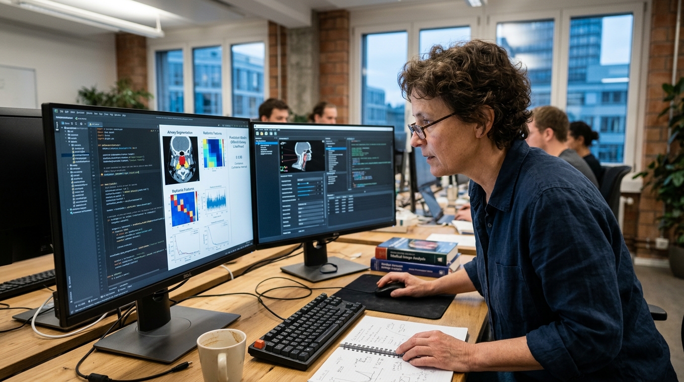
Radiomics is an emerging field that converts medical images (such as Computed Tomography - CT) into mineable, quantitative data. Using advanced algorithms, it extracts features related to the shape, texture, and intensity of tissues within a specific region of interest. In the context of the airway, radiomic analysis focuses on the anatomical structures of the pharynx, larynx, and surrounding soft tissues.
"Radiomics allows us to see beyond anatomy. It enables us to understand the mathematical fabric of tissues and how that fabric dictates airway behavior under anesthesia," the research team notes.
In the recent study, researchers utilized CT scans of patients to train machine learning models. These algorithms learned to identify subtle patterns associated with intubation difficulty. The result was a predictive model with accuracy levels that far exceed any clinical scale currently in use.
Key Findings and Clinical Implications
The findings indicate that AI can predict a difficult airway with an Area Under the Curve (AUC) reaching 0.85-0.90, compared to the meager 0.60-0.70 of traditional methods. This means anesthesiologists can now know with high confidence which patients will require specialized equipment, such as video laryngoscopes or fiberoptic scopes, before the patient even enters the operating theater.
- Personalized Strategy: Each patient is managed based on their unique digital anatomical footprint, reducing trial and error.
- Reduction in Complications: Early preparation minimizes airway trauma and periods of hypoxia.
- Resource Optimization: Hospitals can allocate specialized equipment and senior staff where they are truly needed, improving workflow efficiency.
Challenges and the Future of Digital Anesthesiology
Despite the impressive potential, implementing AI and radiomics into daily practice is not without hurdles. First, there is the issue of cost and availability. Not every patient undergoes a neck CT scan before surgery. However, for patients who already have such imaging for other reasons (e.g., oncology or trauma patients), AI analysis can be performed instantly without additional radiation exposure.
Furthermore, there is the factor of trust. Clinicians must be trained to interpret AI predictions not as absolute truths but as powerful decision-support tools. Ethical questions regarding liability in the event of an AI-related error also remain at the forefront of policy discussions.
In conclusion, the integration of radiomics and AI into anesthesiology represents a massive leap toward precision medicine. As algorithms continue to improve and incorporate data from ultrasound—which is more readily available—the future promises a surgical environment where the "surprise" of a difficult airway becomes a relic of the past. Technology is not replacing the anesthesiologist; it is providing the enhanced vision necessary to protect patient lives with unprecedented precision.