In the delicate ecosystem of Neonatal Intensive Care Units (NICUs), time is both a critical ally and a relentless adversary. For infants born prematurely, the first weeks of life are a continuous struggle to stabilize vital functions. However, a new scientific breakthrough promises to provide doctors and parents with an unexpected tool: a glimpse into the future of a child's brain development, through their very eyes.
According to a recent study highlighted by Fortune Greece, a simple, non-invasive retinal scan could act as a "biological mirror" of the brain. The research indicates that the structure and development of retinal layers in preterm infants are directly linked to brain growth and the risk of neurodevelopmental difficulties, including characteristics within the autism spectrum.
The Biological Connection: Eye and Brain
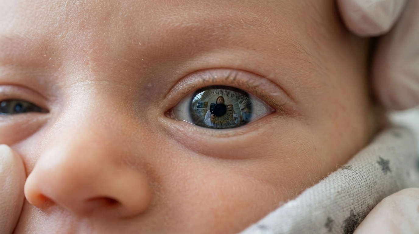
To understand the significance of this study, one must look at embryology. The retina is not merely a light sensor; biologically, it is an extension of the central nervous system. During embryonic development, the retina forms from the same neural tissue that creates the brain. This means that vascular and neural changes observed at the back of the eye often reflect similar processes occurring deep within the cerebral hemispheres.
The study utilized advanced imaging technology known as Optical Coherence Tomography (OCT), which allows for high-resolution images of retinal layers without the need for invasive procedures or radiation. Researchers found that infants with thinner retinal nerve fiber layers or delayed vascularization had significantly higher odds of scoring lower on cognitive and motor tests during toddlerhood.
Predicting Autism and Early Intervention
One of the most striking findings of the research is the scan's ability to identify early markers associated with autism. Until now, autism diagnosis has relied heavily on behavioral observation, which typically does not become apparent until the age of two or three. The ability to identify biological markers as early as the first days or weeks of life radically changes the landscape.
Early diagnosis is not just about prognosis; it is primarily about intervention. The infant brain possesses an extraordinary quality called neuroplasticity. If clinicians know in advance that an infant is at high risk for developmental delays, they can initiate specialized therapies and stimulation programs much earlier, taking advantage of the period when the brain is most receptive to change. This can dramatically improve the child's long-term quality of life.
Ethical Dilemmas and the Future of Neonatology
Despite the excitement, the introduction of such predictive tools brings serious ethical questions. How will parents manage the information that their newborn has an increased probability of autism? Is there a risk of "stigmatizing" the infant before they even begin to interact with the world? Experts emphasize that this technology should be used as a support tool rather than a definitive "verdict."
Furthermore, the issue of access arises. While major university clinics in developed nations possess the necessary equipment, the challenge remains integrating these scans into standard care protocols globally. Reducing the cost of portable OCT devices and utilizing artificial intelligence for automated image analysis could make the exam accessible in every corner of the world.
In conclusion, this study opens a new path in preventive medicine. The fact that we can "read" brain health through the eyes of a preterm infant is a miracle of modern science that reminds us of the profound unity of the human body. The challenge now shifts from the laboratory to clinical practice, with the goal that every child, regardless of how early they entered the world, has the best possible start in life.