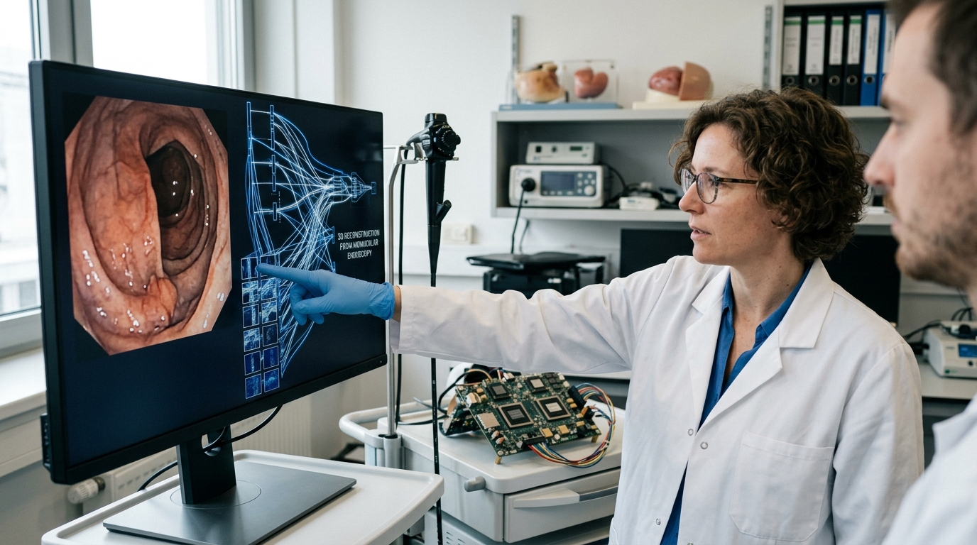
Endoscopic surgery is a cornerstone of modern medicine, enabling procedures with minimal trauma. However, the greatest challenge for surgeons remains the loss of three-dimensional perception. As they look at a 2D monitor to navigate the interior of the human body, the lack of depth can lead to orientation errors or delays. A new pilot study published in the scientific journal Cureus is set to change the status quo, utilizing Artificial Intelligence to convert monocular (2D) images into three-dimensional (3D) views in real-time.
The Challenge of Spatial Perception in the OR
In traditional laparoscopic and endoscopic surgery, the physician works in an environment where the "third dimension" is essentially an illusion created by their experience. The lack of stereoscopic vision—the brain's ability to synthesize images from two different angles to perceive depth—is a significant hurdle, especially for novice surgeons. While 3D endoscopes exist on the market, their cost is prohibitive for many hospitals, and their bulk and technical requirements (such as special glasses) limit widespread adoption.
The study featured in Cureus focuses on "Monocular Depth Estimation" (MDE) via Deep Learning algorithms. This technology does not require new hardware; instead, it "trains" software to recognize shadows, textures, and perspectives within the body, creating a depth map from a standard single-lens camera.
Study Methodology and Results
Researchers conducted a series of tests using simulators and phantom models, comparing the performance of surgeons using standard 2D imagery versus AI-enhanced 3D imagery. The results were striking. The use of AI-driven conversion significantly improved the completion time of delicate surgical maneuvers, such as suturing and object transfer, while reducing error rates during tissue contact.
- Movement Precision: Participants showed 25% greater accuracy in tool placement.
- Time Reduction: Overall procedure times decreased by an average of 15-20%.
- Learning Curve: Trainee surgeons adapted much faster to depth requirements compared to traditional methods.
The most significant finding is that AI managed to provide a sense of depth approaching that of expensive stereoscopic systems, using only the existing equipment already found in hospitals.
The Future: Democratizing High-End Technology
The significance of this research extends far beyond the laboratory. If AI-driven conversion proves stable in broader clinical trials, it could lead to a "democratization" of advanced surgery. Hospitals in developing nations or smaller regional units, which lack the budget for multi-million dollar robotic systems, could upgrade their services simply through a software update.
"Integrating artificial intelligence into endoscopic vision is not just a technical improvement; it is a paradigm shift that transforms raw data into biological perception," the researchers state.
However, challenges remain. Latency in image transmission must be near-zero, as even a few milliseconds of delay can disorient a surgeon. Furthermore, the AI must be trained on a vast variety of pathologies to ensure it doesn't "misinterpret" anatomy due to unusual coloring or bleeding.
Conclusions
The Cureus pilot study is the first step toward a future where the operating room is "smart." The ability of Artificial Intelligence to understand space better than the human eye under constrained conditions promises safer surgeries and faster recovery for patients worldwide. The transition from 2D to 3D via code is perhaps the most cost-effective innovation in medical technology in the last decade.