The era of precision medicine is entering a new, more sophisticated phase as Artificial Intelligence (AI) begins to decode not just 'what' is happening inside a tumor, but also 'where' it is happening. A recent study published in the journal Nature introduces an innovative spatially interpretable AI framework that promises to revolutionize the management of HER2-positive breast cancer. The research focuses on neoadjuvant dual HER2 blockade, a therapeutic strategy administered before surgery to shrink tumors and eliminate micrometastatic disease.
The Critical Role of Spatial Analysis in Oncology
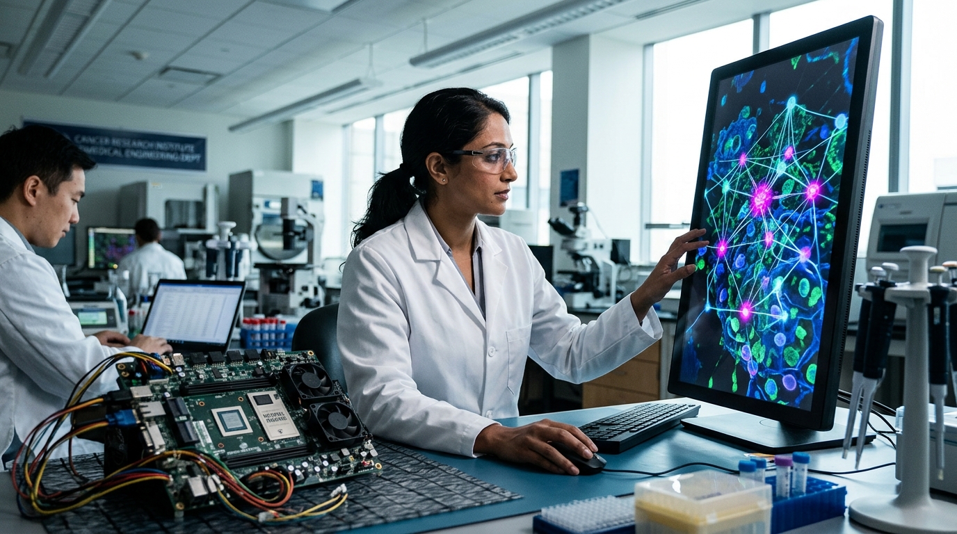
Until now, the evaluation of biopsies has largely relied on the expertise of pathologists and static data regarding the presence of specific proteins or genes. However, cancer is not a homogeneous mass of cells; it is a dynamic ecosystem. The new AI model presented in Nature goes beyond simple cell recognition. It analyzes the spatial organization of the tumor microenvironment (TME), examining how cancer cells interact with immune cells and the surrounding stroma.
This 'spatial interpretability' allows scientists to understand why some patients respond spectacularly to dual therapy with Trastuzumab and Pertuzumab, while others exhibit resistance. The model was trained to identify specific 'neighborhood' patterns within the tissue, which serve as biomarkers to predict pathological complete response (pCR)—the state where no active cancer is detected at the time of surgery.
Personalizing Therapy and Minimizing Toxicity
One of the biggest challenges in modern oncology is treatment 'de-escalation.' Many patients with HER2-positive cancer undergo aggressive chemotherapy regimens that cause severe side effects, even though they might have achieved the same outcome with less toxic means. The AI framework developed can identify with high precision the group of patients who will benefit most from dual HER2 blockade without the need for additional, heavy chemotherapy.
Conversely, for patients whom the model predicts a low probability of response, clinicians can intervene earlier, modifying the treatment plan or introducing novel agents, thus avoiding the loss of valuable time. This approach not only saves resources for the healthcare system but also drastically improves patients' quality of life by protecting them from unnecessary physical and psychological strain.
Toward a New Era of Digital Pathology
Implementing this framework requires the full digitalization of pathology laboratories. Using deep learning algorithms in combination with spatial transcriptomics technologies allows for the mapping of gene expression directly onto tissue sections. The fact that this specific model is 'interpretable' is key to its adoption by the medical community. Unlike the 'black boxes' of traditional algorithms, this system explains to clinicians which specific tissue features led to the prediction, fostering trust in clinical decision-making.
In conclusion, the Nature study marks a transition from general statistics to personalized spatial analysis. As AI becomes more refined, the ability to 'read' the geography of cancer will become an indispensable tool for every oncologist, transforming diagnosis into a precision roadmap for treatment. The integration of spatial AI into clinical workflows signifies a major step toward truly individualized cancer care, where the biological uniqueness of each patient dictates the therapeutic path.