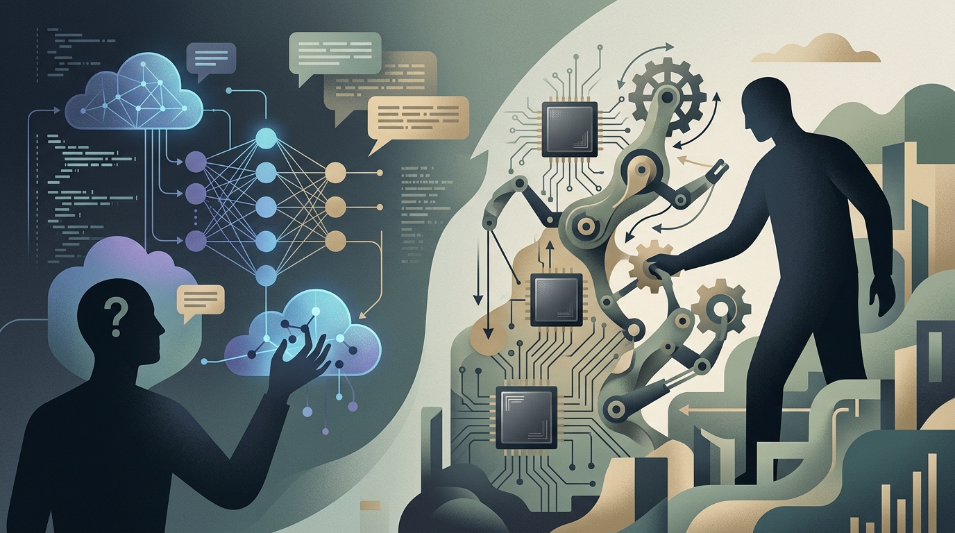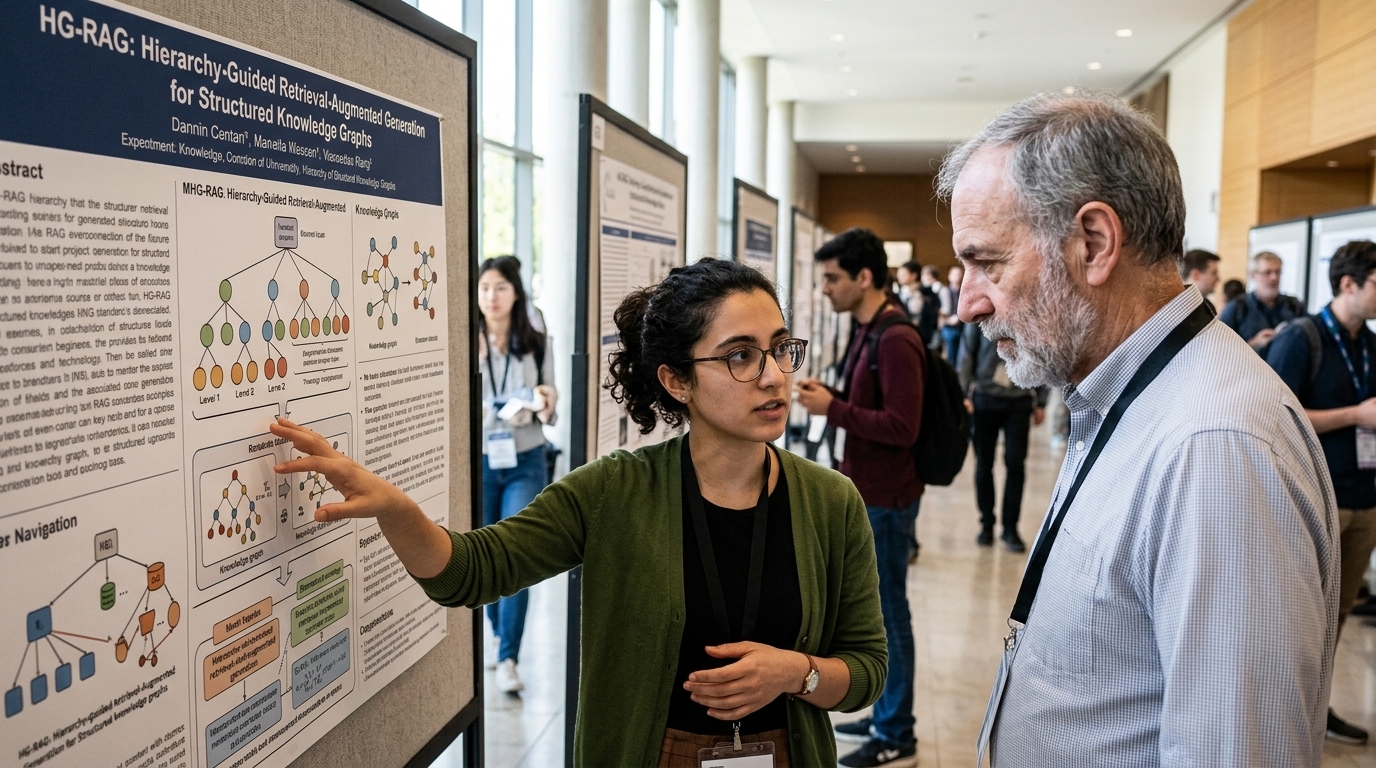
For decades, Computed Tomography (CT) has been one of modern medicine's most powerful yet controversial tools. On one hand, its ability to "slice" through the human body and detect malignancies at an early stage has saved millions of lives. On the other, the ionizing radiation required to capture these high-resolution images has always carried a small but tangible risk of inducing secondary cancers. Today, in the summer of 2026, Artificial Intelligence (AI) appears to be providing the definitive solution to this medical dilemma, radically transforming our approach to preventative screening.
The Radiation Paradox and the Digital Solution
The fundamental challenge in CT imaging has always been the signal-to-noise ratio. To obtain a clear image that allows for the diagnosis of microscopic tumors, radiologists had to use high doses of radiation. Reducing the dose inevitably led to "blurry" or "grainy" images, rendering them useless for clinical use. This problem was particularly acute in lung cancer screening, where frequent repeat examinations accumulated dangerous levels of radiation in patients' bodies over time.
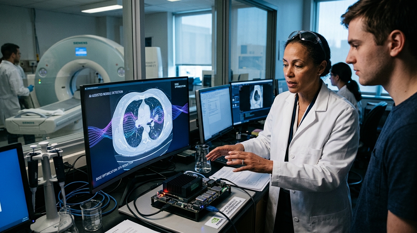
The advent of Deep Learning and image reconstruction algorithms has changed the landscape. New AI algorithms, trained on millions of pairs of low-dose and high-dose images, can now "predict" and fill in the missing details from an ultra-low-dose scan. The result is an image that looks like a traditional high-dose CT but is captured with as little as 1/10th of the radiation. This development is not merely a technical improvement; it is a paradigm shift in public health.
Application in Practice: From Vietnam to the Global Community
Recent reports from medical centers in Vietnam, which are rapidly adopting these technologies, show that the use of AI in cancer screening allows for the expansion of screening programs to population groups previously considered "high risk" for the procedure itself. In Europe and beyond, the integration of these systems into public hospitals is accelerating, as the reduction in radiation risk makes CT scans nearly as safe as a simple chest X-ray.
Furthermore, AI is not limited to image denoising. Modern platforms feature Computer-Aided Detection (CAD) systems that point out areas of interest to the radiologist that the human eye might miss due to fatigue or anatomical complexity. This human-machine collaboration reduces false negatives, ensuring that cancer does not go undetected during the window of opportunity for treatment.
Ethical Challenges and the Future of Diagnosis
Despite the excitement, the use of AI in medical imaging raises significant questions. One is the "illusion of accuracy." As AI "fills in" the gaps in an image, there is a risk of creating artifacts that do not exist in reality or, conversely, smoothing out pathological lesions by mistaking them for "noise." Regulating these systems requires strict protocols and continuous calibration to ensure patient safety remains paramount.
- Personalized Dosing: In the near future, AI will calculate the exact radiation dose based on each patient's body type and genetic profile.
- Accessibility: Reducing costs and risks will allow for the use of mobile CT units in remote or underserved areas.
- Data Integration: CT images will be automatically combined with blood biomarkers for a holistic, multi-modal diagnosis.
In conclusion, Artificial Intelligence is not replacing the radiologist; it is providing them with a "super-weapon." Eliminating the fear of radiation clears the path for a new era in preventative medicine, where cancer is no longer a death sentence but a manageable condition detected long before symptoms even appear.